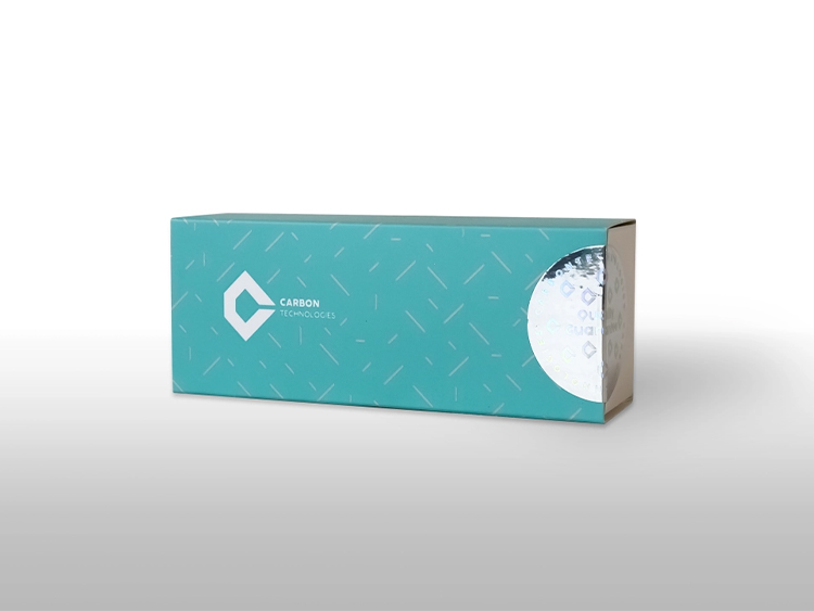
Stark MTB Molecular Diagnostic Kit
Catalog Number: ST242002
Package Specification: 25 tests/kit
Kit Content
Ingridients | 25 Preps | 100 Preps |
Pro MTB Mix | 250 µl | 1000 µl |
QR-ROMAX, 4X | 125 µl | 500 µl |
MTB Positive Control | 100 µl | 150 µl |
RT-PCR Grade Water | 100 µl | 1500 µl |
Intended Use
The Stark MTB Molecular Diagnostic Kit Real-Time PCR Kit is intended for in vitro diagnostic use by trained healthcare professionals. Designed for the detection of Mycobacterium tuberculosis (MTB) DNA in human samples, including sputum, bronchoalveolar lavage (BAL), bronchial secretion, cerebrospinal fluid (CSF), stomach fluid, peritoneal puncture, and urine, this kit employs Real-Time PCR technology for precise and reliable results. The obtained data aids healthcare professionals in diagnosing MTB complex infections, guiding treatment decisions, and monitoring patient response. Strict adherence to the provided instructions is imperative to ensure accurate interpretation of results and effective utilization of this diagnostic tool.
Application
The Stark MTB Molecular Diagnostic Kit is an in vitro nucleic acid amplification test designed for the detection of all members of the M. tuberculosis complex (M. tuberculosis, M. africanum, M. bovis, M. bovis BCG, M. microti, M. pinnipedii) in human sputum, bronchoalveolar lavage (BAL), bronchial secretion, cerebrospinal fluid (CSF), stomach fluid, or peritoneal puncture samples. This diagnostic test kit utilizes polymerase chain reaction (PCR) technology and is compatible with Real-Time PCR instruments.
Sample Collection and Preparation
Materials Required (but Not Provided)
- DNase-RNase-free microtubes (1.5 ml)
- PCR microtube 0.1 ml or 0.2 ml strip
- Various models of pipettes and pipette tips (10 µl, 100 µl, and 1000 µl of filter pipette tips)
- Surface sanitizing solution such as RNZO
- Disposable Powder-Free gloves and surgical gown
- Different types of Real-Time PCR Instruments (with green, yellow channels)
- Centrifuge (capable of reaching 13000 rpm)
- Microcentrifuge
- Cool box
Procedures
Tuberculosis is an airborne mycobacterial infection caused by the M. tuberculosis complex (M. tuberculosis, M. africanum, M. bovis, M. bovis BCG, M. microtia, M. pinnipedii). MTBC is transmitted from one person to another through tiny droplets released into the air via coughs and sneezes. Early detection of TB is paramount to improving health outcomes for individuals with TB and effectively reducing TB transmission. The Stark MTB Molecular Diagnostic Kit employs a polymerase chain reaction-based Real-Time PCR technique. Specifically designed for the qualitative diagnosis of the IS6110 gene (a specific multi-copy insertion sequence) of Mycobacterium tuberculosis, this kit enables precise detection. After nucleic acid isolation using the DNJia Tissue and Bacteria Kit or other Ministry of Health-approved kits, the verified sample combination is added to the master mix primer/probe mix for the reaction. Furthermore, the Stark MTB Molecular Diagnostic Kit incorporates a secondary heterologous amplification system to identify potential PCR inhibition. This is detected as an internal control (IC) in the fluorescence channel Cycling Yellow of the Rotor-Gene Q MDx, Rotor-Gene Q, or Rotor-Gene 6000, or Cycling A.JOE of the Rotor-Gene 3000. Through its sampling mechanism, the quality of sample isolation and PCR reaction process can be monitored and controlled to prevent false-negative results. The assay demonstrates a Limit of Detection (LoD) of 25 Copies/ml.
Applications
The Stark MTB Molecular Diagnostic Kit is an in vitro nucleic acid amplification test designed for the detection of all members of the M. tuberculosis complex (M. tuberculosis, M. africanum, M. bovis, M. bovis BCG, M. microti, M. pinnipedii) in human sputum, bronchoalveolar lavage (BAL), bronchial secretion, cerebrospinal fluid (CSF), stomach fluid, or peritoneal puncture samples. This diagnostic test kit utilizes polymerase chain reaction (PCR) technology and is compatible with Real-Time PCR instruments.
Recommended Starting Material
Before initiating any tests, ensure that each component is thawed by gently bringing it to room temperature. Perform gentle up and down mixing, followed by a brief spin and centrifugation. Avoid subjecting the components to repeated freeze-thaw cycles.
Sample Storage and Preparation
For human sputum, bronchoalveolar lavage (BAL), bronchial secretion, cerebrospinal fluid (CSF), stomach fluid, or peritoneal puncture samples, the following guidelines apply:
Fresh specimens should either be processed immediately according to the sample procedure outlined in the Sample Processing Protocol section or stored frozen at -20°C. Prior to starting sample processing, frozen samples must be allowed to thaw to room temperature. Sample pre-treatment involves decontamination of the specimen, preparing it for extraction.
Before Starting
Remove each component from the kit and place them on the benchtop. Allow the reagents to equilibrate to room temperature. Then, gently mix each tube by performing up and down motions and briefly spin each tube in preparation for later use.
Buffer Preparation
Table: Outlines the preparation of components per single reaction.
Volume | Components |
5µl | QR-ROMAX, 4X |
10µl | Pro MTB Mix |
5μl | Isolated DNA |
Pathogenicity
Mycobacterium tuberculosis is a species of pathogenic bacteria belonging to the family Mycobacteriaceae and is the causative agent of tuberculosis. It was first discovered in 1882 by Robert Koch. According to the most recent Global Tuberculosis Report (2019) edited by the World Health Organization (WHO), tuberculosis is ranked as the ninth leading cause of death worldwide and is the primary cause of mortality by a single infectious agent, with the highest rates of infection and mortality predominantly observed in developing and low-income countries.
Human infections with MTB typically begin through the inhalation of aerosol droplets containing tubercle bacilli directly expectorated from individuals with “open” pulmonary disease. The infectious dose for an individual is estimated to be between 1 and 200 bacilli; however, as a single aerosol droplet can contain anywhere from 1 to 400 bacilli, it remains uncertain what constitutes a biologically relevant dose. Following inhalation, the bacilli travel to the alveoli, where they are rapidly phagocytosed by alveolar macrophages.
The pathogenicity of MTB primarily stems from its ability to reprogram host macrophages after primary infection, thereby evading its own elimination. This includes the formation of granulomas, wherein the pathogen survives in equilibrium with host defenses, as well as the modulation of bacterial central metabolism and replication, leading to a dormant state wherein MTB becomes resistant to both host defenses and therapy.
Workstation Preparation
Before commencing work, ensure that all work surfaces, pipettes, centrifuges, and other supplies are thoroughly cleaned and sanitized. To minimize the risk of nucleic acid contamination, utilize sanitizers such as 70% Ethanol or 10% Sodium Hypochlorite.
Protocols
Thaw all reagents completely at room temperature (15–25°C). Once thawed, thoroughly mix all reagents by gently performing up and down motions, followed by brief spinning and centrifugation. Work promptly and maintain all reagents in the cooling block to preserve their integrity.
PCR Reaction Preparation
Table : PCR Reaction Preparation
Volume | Components |
5µl | QR-ROMAX, 4X |
10µl | Pro MTB Mix |
5μl | Isolated DNA |
Thermal Profile
Table : Thermal profile for PCR reaction
Stage | Temperature | Incubation Time | Cycle Numbers |
Pre-Denaturation | 95°C | 3 min | 1 |
Denaturation | 95°C | 10 sec | 45 |
Annealing and acquisition on channels Green and Yellow | 60°C | 40 sec |
Acceptable Situations for Positive and Negative Control
Table : Control conditions for a valid PCR run
Results | IC (HEX) | MTB (FAM) |
Positive control* | Not considered | Ct≤40 (+) |
Negative control | Ct>35 (+) | – |
Invalid and not accepted | – | – |
Quality Control
The Stark MTB Molecular Diagnostic Kit undergoes rigorous testing in accordance with clinical, laboratory standards, and guidelines set forth by institutes such as the WHO. These tests are conducted through predefined experiments on a lot-to-lot basis to uphold consistent product quality. For your convenience, detailed results of all experiments can be accessed online by referencing the REF and Lot numbers at www.amorph.tech.
Results
- Perform data analysis for each gene separately using a manual threshold.
- Ensure that the threshold for each sample is set within the exponential phase of the fluorescence curves and above any background signal.
- Utilize the FAM Fluorophore (green) for the IS6110 gene of M. tuberculosis and the HEX Fluorophore (Yellow) for the internal control (IC) gene.
- Employ a negative control to monitor contamination. Any significant increase in fluorescence curve magnitude in the negative control that does not cross the threshold, with a Ct value less than 35 (Ct<35), may indicate possible contamination. Strong signals above 35 in the negative control can suggest PCR artifacts; in such cases, consider the curve shape (an S-shaped curve typically indicates a positive result).
- Ensure that the internal control yields a positive result for all clinical specimens with a Ct value of 35 or less, indicating sufficient nucleic acid from the human gene and acceptable sample quality.
- A Ct value greater than 33 or an absence of Ct for the internal control indicates low sample concentration or inhibitors in the reaction. In such instances, it is recommended to dilute the isolated sample by at least half. If the test result remains unacceptable upon retesting, obtain a new sample from the patient and repeat the test.
- Positive clinical specimens should have a Ct value of ≤40 for the target gene (IS6110).
- If the expected positive reaction, characterized by a typical S-shaped curve, is not achieved, the performed test is deemed unacceptable and must be repeated in accordance with the kit instructions.
- Identify the cause of the positive control failure, implement corrective actions, and document the results of corrective actions.
Specific Characteristics
Real Time-PCR | Technology |
qualitative | Type of Analysis |
IS6110 genes (specific multi-copy insertion sequence) | Target Sequence |
Mycobacterium tuberculosis complex (M. tuberculosis, M. bovis, M. africanum, M. microti, M. caprae, M. canetti and vaccine strain BCG), 100% | Analytical Specificity |
LOD of assay is 25 Copies/ml with the probability 95% | Analytical Sensitivity (LOD) |
100% (CI95%: 99.06% –100%) | Diagnostic Specificity |
100% (CI95%: 99.06% –100%) | Diagnostic Sensitivity |
PCR inhibition and DNA extraction efficiency control | Extraction/Inhibition Control |
Human sputum, BAL, bronchial secretion, CSF, stomach fluid or peritoneal punction | Validated Specimen |
-20 ± 5°C | Storage |
DNJia Tissue and Bacteria Kit | Validated Extraction Method |
Rotor-Gene Q, 2plex, Corbett Rotor-Gene 3000&6000, Mic qPCR Cycler, StepOne and StepOne plus Applied Biosystem | Instruments |
Green-Yellow | Required Detection Channels |
For bellow information refer to the Product IFU:
- Limit of Detection (LoD) – Analytical Sensitivity
- Clinical Sensitivity
- Cross-reactivity (Analytical Specificity)
- Cross-reactivity (Clinical Specificity)
- Precision Assessment
- Intra-assay
- Inter-assay
- Clinical Evaluation
Limitation of the Procedure
- A false-negative result may occur due to low titration of MTB in the patient sample, improper transportation, or inadequate sample isolation quality.
- Verification of all controls is essential before interpreting the results. If the controls are invalid, the patient’s results cannot be accurately interpreted. The diagnostic threshold of this kit is Ct≤40, and the user must review the fluorescence curve before making a final interpretation. All positive curves should exhibit an amplification peak.
- Failure to observe proper storage conditions for the kit may lead to false-negative results.
- Handling of this kit requires experienced and trained personnel. Any errors made by personnel may result in invalid results.
- The results obtained from this diagnostic kit are considered acceptable only when supported by clinical evidence for diagnosing MTB. Definitive diagnosis and treatment decisions for patients should be based on a combination of this test with other test results, medical records, and the patient’s response to treatment.
Storage and Safety
Storage
All components of the Stark MTB Molecular Diagnostic Kit are pre-prepared and ready for immediate use upon arrival. Upon receipt, it is recommended to store all reagents at temperatures ranging from -15°C to -30°C. These conditions ensure stability and maintain the integrity of the components until the expiration date indicated on the label.
Warning and Precautions
- Material Safety Data Sheets (MSDS) for all products and reagents are available online at http://www.amorph.tech. Users are advised to adhere to laboratory safety protocols diligently.
- Please review the guidelines thoroughly before initiating use.
- All patient samples and positive controls should be treated as potentially infectious.
- Consumption of food, beverages, tobacco, gum, cosmetics, or medication is strictly prohibited in laboratories handling hazardous materials and human samples. All patient samples and positive controls must be regarded as potentially infectious.
- The Stark MTB Molecular Diagnostic Kit is intended for emergency and in vitro diagnostic use as directed by a physician’s prescription.
- Each procedural step, including sampling, storage, shipping, and laboratory testing, must comply with biosafety and molecular laboratory management protocols.
- The Stark MTB Molecular Diagnostic Kit requires a dedicated and private laboratory space:
- Location 1: Preparation Area – for test component preparation.
- Location 2: Sample Processing Area – for isolation and control.
- Location 3: Amplification Area – for Real-Time PCR testing.
- Clinical laboratories must be equipped with instruments and personnel compliant with Ministry of Health regulations.
- Altering or substituting any kit components may impair its function and void the product license.
- All pipette tips and microtubes must be sterile and free from DNase and RNase contamination. To prevent cross-contamination, filter pipette tips should be used and replaced after each substance or sample addition.
- Dispose of waste in accordance with biosafety guidelines. Desks and laboratory instruments should be regularly disinfected with 70% ethanol or 10% sodium hypochlorite.
- Protect the Pro MTB combination from sunlight exposure.