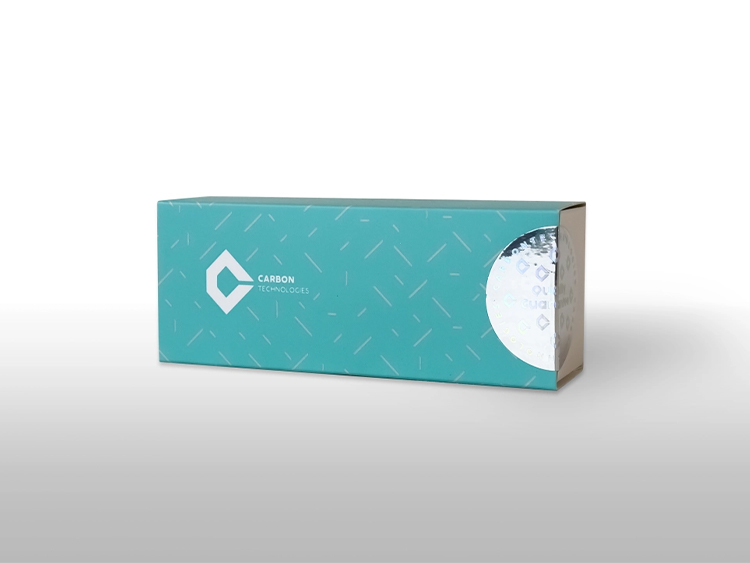
Stark SARS-CoV-2 & Influenza A/B Molecular Diagnostic Kit
Catalog Number: ST242003
Package Specification: 100 tests/kit
Kit Content
Components | 100 Preps |
Q-ROMAX, 4X | 500µl |
Pro II Mix | 400µl |
RTase, Recombinant Reverse Transcriptase, RNase H-(200 U/µl) | 100µl |
Positive Control | 150µl |
Negative control | 150µl |
Intended Use
The Stark SARS-CoV-2 and Influenza A/B Molecular Diagnostic Kit is designed for accurate detection of the N gene of SARS-CoV-2, the Matrix Protein gene of Influenza A, and the NS1 gene of Influenza B in individuals suspected of infection. Suitable samples include those obtained from the upper and lower respiratory tracts, such as nasopharyngeal swabs, throat swabs, nasal washes, nasal aspirations, sputum, and bronchoalveolar lavage. These samples may be collected from patients exhibiting symptoms indicative of COVID-19 or influenza or from healthcare personnel potentially exposed to these viruses. The kit’s comprehensive approach ensures accurate and reliable detection, aiding healthcare providers in timely diagnosis and appropriate management of patients.
Application
The Stark SARS-CoV-2 and Influenza A/B Molecular Diagnostic Kit utilizes a one-step RT-PCR reverse transcriptase reaction to detect specific RNA sequences from pathogens. This process involves generating complementary DNA (cDNA) from the RNA template of the pathogen, followed by PCR amplification. The resulting PCR products are then identified using oligonucleotide probes labeled with fluorescent colors. This kit is designed to detect the N gene from SARS-COV-2 (Coronavirus), the M2 gene from Influenza A, and the NS1 gene from Influenza B virus. Other Coronaviruses and strains of the Influenza virus are not detected by this kit.
The primer/probe sets for the N gene of COVID-19, the M gene of Influenza A, and the NS1 gene of Influenza B are designed to target conserved regions among their respective pathogens. These genes are detected in specific fluorescence channels: the N gene in the orange channel, the M gene in the green channel, and the NS1 gene in the yellow channel. The internal control, RNase P gene, is detected in the Cy5 channel, serving as a control to isolate RNA and check for RT-PCR reaction inhibition. This kit is compatible with Real-Time PCR devices capable of identifying four fluorescence channels (green, yellow, orange, and red).
Sample Collection and Preparation
Materials Required (but Not Provided)
Here are the materials required for the STARK SARS-COV-2 Molecular Diagnostic Kit, which are not provided:
- Nylon or Dacron swab with an aluminum or plastic shaft for sampling.
- DNase-RNase-free microtubes (1.5ml).
- PCR microtube 0.2- or 0.1-ml strip.
- Various pipettes and pipette tips (10µl, 100µl, and 1000µl of filter pipette tips).
- Surface sanitizing solution.
- Disposable Powder-Free gloves and surgical gown.
- Different types of Real-Time PCR Instruments (green, yellow, orange, and red channels).
- Centrifuge (which can reach 13000 rpm).
- Microcentrifuge.
- Vortex.
- Cool box.
Real-Time PCR Instruments
The STARK SARS-COV-2 Molecular Diagnostic Kit is compatible with the following Real-Time PCR devices:
- Rotor-Gene Q, 5plex
- Corbett Rotor-Gene 3000 & 6000
- Mic qPCR Cycler
- ABI Step One & Step One Plus
- Biorad CFX96 Real-Time PCR
- Roche LightCycler® 96 Real-Time PCR System
- Anatolia Montania 484 Real-Time PCR Instrument
Please note that some PCR devices may require calibration with the desired colors before performing multiplex-PCR reactions. For further details about specific Real-Time PCR devices, refer to the device manual template.
Sample Collection
All samples must be collected to avoid contamination during sampling, storage, and transportation. Samples should be treated as potentially infectious, following biosafety guidelines. Synthetic-tipped swabs with aluminum or plastic shafts are recommended for collection, while cotton swabs with wooden shafts should be avoided. After sampling, swabs should be stored immediately in a virus transport medium.
Storage and Delivery of Specimens
Samples should be tested within 24 hours if stored at 4ºC. For longer storage, samples can be stored at -70ºC or below. If -70ºC storage is unavailable, specimens can be stored at -20ºC for up to ten days, and nucleic acid can be stored at -20±5ºC for 15 days. Avoid multiple freeze-thaw cycles to maintain sample integrity.
Specimen Isolation
For viral nucleic acid isolation, use a kit which is approved by the Ministry of Health.
Pathogenicity
Coronaviruses belong to the Coronaviridae family, characterized by positive single-stranded RNA genomes. Strains such as HCOV-229E, HCOV-NL63, HCOV-OC43, MERS-CoV, and HCOV-HKU1 are known to cause various respiratory illnesses in humans, including the common cold, upper respiratory tract infections, bronchiolitis, and pneumonia. SARS-CoV-2, identified as a beta coronavirus in December 2019 in Wuhan, China, is the causative agent of COVID-19. Fever, cough, and respiratory issues are among the primary symptoms, often progressing to pneumonia and severe respiratory syndrome (SARS). The virus primarily spreads through close contact with respiratory droplets.
Influenza is an acute infectious disease caused by Influenza A and B viruses, with Influenza C viruses causing fewer cases. The genome of influenza viruses comprises segmented RNA strands encased in a protein capsid with a negative head. These viruses are ubiquitous globally. The spread of influenza A is primarily attributed to antigenic drift of hemagglutinin and neuraminidase molecules. While Influenza B and C viruses are predominantly human pathogens, Influenza A viruses have a broader host range, infecting various warm-blooded animals.
Workstation Preparation
Before commencing any procedures, it is essential to prepare the workstation diligently. Thorough cleaning and sanitization of all work surfaces, pipettes, centrifuges, and other equipment are imperative. To minimize the risk of nucleic acid contamination, utilize sanitizers such as 70% Ethanol or 10% Sodium Hypochlorite. This ensures a clean and safe environment conducive to accurate testing procedures.
Process
Retrieve each component from the diagnostic kit and set them aside at room temperature. Let the reagents equilibrate to room temperature before proceeding, and then briefly vortex each to ensure proper mixing for later use. Ensure that the volume of the isolated sample used in this test is 10µl. Refer to table 1 to prepare the reaction components accordingly and conduct Real-Time PCR as outlined in table 2.
Table 1: Preparation of reaction components for one reaction.
Components | Volume |
Q-ROMAX | 5µl |
RTase, Recombinant Reverse Transcriptase, RNase H-(200 U/µl) | 1µl |
Pro II Mix | 4µl |
Isolated RNA | 10µl |
Table 2: Temperature program of one-step Multiple Real Time-RT-PCR
Step | Time | Temperature | Number of cycles |
cDNA synthesis | 20min | 50ºC | 1 |
Polymerase enzyme activation | 3min | 95ºC | 1 |
Denaturation | 10s | 95ºC | 45 cycles |
Annealing and extension of nucleic acid and measurement of fluorescence in green, yellow, and orange channels | 40s | 60ºC |
Protocol
Quality Control
In adherence to standards set by the Clinical and Laboratory Standards Institute and WHO, the STARK SARS-COV-2 Molecular Diagnostic Kit undergoes rigorous testing across multiple experiments on a lot-to-lot basis to guarantee consistent product quality. Detailed results of these experiments are accessible online by referencing the REF and Lot numbers at www.carbontechnoloesco.com.
Results
- Data analysis for each gene should be performed separately using a manual threshold.
- The threshold for each sample should be in the exponential phase of the fluorescence curves and above any background signal.
- FAM Fluorophore (green) to detect Influenza A, Yakima Yellow Fluorophore (Yellow) to detect Influenza B, Texas Red Fluorophore (orange) to detect COVID-19, and Cy5 Fluorophore (Red) is for the RNase P gene (internal control).
- A negative control is used as contamination control. The magnitude increases of the fluorescence curve in the negative control do not cross the threshold. If Ct is less than 35 (Ct<35), it is considered possible contamination. Strong signals above 35 in the NTC can be PCR artifacts which, in these cases, the shape of the curve can be considered (the S-shaped curve is typical for a positive result).
- Internal control or RNase P gene should be positive for all clinical specimens at Ct 35 or less than 35, indicating sufficient nucleic acid from the human RNase gene and the sample has acceptable quality.
- Internal control curve or RNase P gene Ct>40 or without Ct indicates low sample concentration or inhibitors in the reaction (recommended that the isolated sample be diluted at least ½). If the test result is not acceptable again during the retest, another new sample should be taken from the patient, and the test must be repeated.
- A positive clinical specimen Ct≤40 for the green, yellow and orange channels indicate Influenza A, Influenza B, and COVID-19, respectively. If any of the above channels are positive and the Red channel (internal control) is not, it is considered a negative sample if only the Red channels are positive otherwise, our result is not valid.
- A positive clinical specimen should have Ct≤40 for each of the three genes or positive genes.
- If the expected positive reaction is not achieved (typical S-shaped curve), the performed test is not acceptable, and repeat the test by following the kit instructions exactly.
- Determine the reason for the failure of positive control and the corrective action, and document the corrective action results.
Specific Characteristics
For information about these bellow subjects refer to the product IFU:
- Limit of Detection (LoD) – Analytical Sensitivity
- Clinical Sensitivity
- Cross-reactivity (Analytical Specificity)
- Cross-reactivity (Clinical Specificity)
- Accuracy Assessment
- Intra-assay
- Inter-assay
- Clinical Evaluation
Limitation of the Procedure
The efficacy of this test is contingent upon the collection, handling, and storage of samples. Specifically designed for diagnosing targeted viruses in swab samples and respiratory sputum, this kit performs optimally when utilized on samples obtained from individuals displaying symptoms of COVID-19 or influenza. Samples may be self-collected or obtained by healthcare personnel. A negative test result does not definitively rule out the presence of SARS-CoV-2, Influenza A, or B viruses, as various factors such as sample collection technique, user error, mixing methodology, or low viral titers can potentially impact the accuracy of the test. Additionally, the presence of PCR inhibitors may lead to false-negative results. Moreover, sequence variations in the target regions of unknown viruses may also contribute to false-negative results or reduced kit sensitivity. Therefore, the interpretation of results should be based on clinical assessments and complementary diagnostic tests.
Storage and Safety
Storage
Upon receipt, store all reagents in a dark environment at -20 ±5ºC. Avoid frequent freeze-thaw cycles of the kits. When stored according to the specified conditions, all reagents remain stable until the expiration date indicated on the kit box.
Warning and Precautions
- Ensure that only personnel who are specially trained and instructed in handling RT-PCR diagnostic cases utilize this product.
- Regular maintenance and repair of the Real-Time PCR instrument are necessary.
- Clean tables and equipment periodically to maintain hygiene standards.
- Utilize sterile filter pipette tips that are free from RNase and DNase.
- Always treat samples as infectious and handle them with care according to laboratory safety protocols. Wear powder-free latex gloves when handling kit materials and components.
- Establish separate workstations for sample preparation and reaction setup. Each workstation should be equipped with dedicated tools and equipment, and airflow should be directed from pre-PCR to post-PCR areas.
- Exercise caution when working with samples containing high virus titers and positive controls to prevent laboratory contamination.
- Change gloves after handling samples or positive controls, and keep them segregated from other reaction materials.
- Prevent contamination of workstation materials and equipment with DNA/RNA and nucleases.
- Ensure that the RNA isolation method is compatible with the RT-PCR reverse transcriptase technique to maintain the quality of isolated RNA.
- To prevent false-positive results due to viral RNA contamination during isolation, use a negative control (water instead of sample) during RNA extraction and PCR testing. Optionally, include a negative control sample (water instead of sample) in each PCR reaction.
- Monitor the expiration date of the Kit and avoid repeated freezing and thawing of its components. Store Kit components away from light.
- Dispose samples following laboratory waste disposal safety regulations.